LESSER ORDER VEINS:
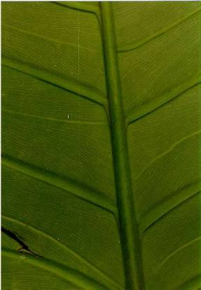
Between successive primary lateral veins there are possibilities for two additional orders of veins. Frequently present are intermediate primary lateral veins called "interprimary veins" (Figs. 24, 26) [See also Croat & Bunting, 1979]. These veins, while decidedly less conspicuous than the primary lateral veins, are nevertheless too prominent to classify as the smallest order veins. To qualify as an interprimary vein the vein must extend continuously from the midrib to very near the margin without major branching. Generally there is no more than one pair of interprimary veins between alternate primary lateral veins. They are akin to the primary lateral veins in all aspects except for their greatly reduced size. Like the primary lateral veins they may bear minor veins which may form all along their margins.
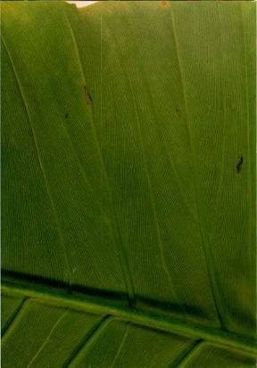
The smallest order veins are referred to as "minor veins" (Fig. 24) and may be close, fine and conspicuous as in P. sulcicaule, P. tripartitum, and P. radiatum, to thick, well-spaced and inconspicuous on P. gigas, P. granulare, P. grayumii, and P. ligulatum. The minor veins are not all equally distinct and sometimes, as in P. dominicalense, the minor veins are alternately weakly visible and strongly visible.
The minor veins may arise from the midrib or from the primary lateral and interprimary veins but in either case they form a generally close, uniform and parallel array which extends without interruption to near the margin of the blade. In most species the minor veins arise from both the midrib and the primary lateral veins but some species have the minor veins arising from only the midrib. A total of 77 species (81 taxa) have the minor veins arising from both the midrib and the primary lateral veins (Fig. 26). In such cases the minor veins are not always equally arising from one of the two entities but may, such as in the case of P. brenesii, P. davidsonii var. davidsonii, P. ferrugineum, and P. tripartitum, be more heavily arising from the midrib rather than the primary lateral veins. In the case of P. auriculatum, P. glanduliferum, P. lentii, and P. ligulatum the minor veins arising from the primary lateral veins are many fewer than those arising from the midrib. In P. heleniae the primary lateral veins are only rarely arising from the midrib.
In another variation of this venation type, some species, while having minor veins arising from the midrib as well as both adjacent primary lateral veins have considerably more veins arising from the primary lateral vein which is more distil than from that vein which is basal, ie. closer to the petiole.
A total of 24 species have the minor veins arising from only the midrib and in this case they course along the primary lateral veins but do not join with it. Many of the species that have the minor veins arising only from the midrib are species with oblong blades including P. bakeri, P. cretosum, P. dolichophyllum, P. granulare, P. heleniae, P. roseospathum, P. ubigantupense, P. utleyanum, and P. wendlandii. However, the group also contains blades which are ovate or nearly so such as P. brewsteriense, P. chirripoense, P. cotobrusense, P. crassispathum, P. folsomii, P. knappiae, P. microstictum, P. niqueanum, P. sulcicaule, and P. verapazense. Interestingly P. anisotomum with deeply 3-lobed leaf blades also has the minor veins arising from only the midrib, whereas P. tripartitum another 3-lobed species with which it may be confused has the minor veins arising from both the midrib and the primary lateral veins.
Philodendron dressleri, a species with deeply divided leaves, has a more complex venation pattern. Although the minor veins arise from both the midrib and the primary lateral veins they also arise from short secondary veins which regularly branch off of the primary latteral veins. In addition the minor veins which arise from the midrib are considerably fewer in number and weaker than in most species with this venation pattern. Generally the confluent minor veins which arise from the primary lateral veins and make a broad sweep before continuing to the margin leave little area for the minor veins which arise from the midrib. Generally the latter merge imperceptably with those from the primary lateral veins. A similar pattern with weak midrib borne minor veins is present with P. basii.
At or very near the margin both the primary lateral veins and the minor veins generally turn sharply upwards toward the apex of the blade. The minor veins merge with other minor veins and finally merge with the primary veins before finally merging into an inconspicuous marginal and somewhat opaque marginal plexus. This narrow band is usually chlorophyllous and apparently veinless. Frequently the outer margin of this chlorophyllous band is a hyaline edge which is colorless and typically revolute.
The minor veins are sometimes noticeably interconnected by inconspicuous to conspicuous veins, referred to here as "cross-veins" (Fig. 26). Generally the cross-veins are markedly perpendicular to consecutive minor veins where they are sufficiently prominent to be noticeable, but in some cases the cross-veins cross transversely from one minor vein to the next. While P. scalarinerve has cross-veins so prominent, even on fresh material, as to be easily visible, other species such as P. chiriquense and P. copense, have the cross-veins which are easily visible only when the blades are dry.
Frequently present on Central American members of P. subg. Philodendron are secretory ducts and other secretory tissues. The secretory ducts may have contents that are either latex or taniniferous compounds (Solereder & Meyer, 1928). No thorough survey of Central American species has been made of the nature of the secretory canals so it is not always apparent whether these structures are resin canals or secretory ducts. Secretory canals (also referred to as secretory files) in Philodendron are always non-anastomosing and consist of a linear sequence of secretion cells, each separated from the next by cell walls (Solereder & Meyer, loc. cit.). On fresh leaves and dried leaves these can usually be determined by being darker, usually blackened and in being intermittent rather than continuous as is generally true of veins. While the distribution of secretory ducts in Philodendron may be more common than is apparent from surface examination, not all species exhibit the secretory ducts clearly. Thus the presence or absence of distinct secretory ducts can be useful taxonomically. They are distinctly visible in P. alticola, P. cotonense, P. grayumii, P. heleniae, and P. zhuanum but obscurely visible on P. antonioanum, and P. bakeri. They are clearly visible on P. schottianum and somewhat visible on P. llanoense and very obscure on P. findens even though these three species have very similar leaf blades.
In addition, the surface between the minor veins is frequently marked by other features, including pale sub-surface granulations (perhaps indicating the presence of druse crystals), short, pale lineations, gland-like punctiform markings, reddish or brownish speckling and also what might be referred to as "stitching", pale intermittent short lines appearing on the surface of the blade as though the blade was loosely sewn with a needle and thread. Though this phenomenon is much more common in P. subg. Pteromischum it is also exhibited in P. subg. Philodendron.
While perhaps no more reliable than blade shape, blade size or other features, blade surfaces at a magnification of 10X or higher often yield another suite of characters, such as those mentioned above, which often yield another degree of confidence (or forewarn of misidentification) when making determinations.